The 'Society for General Microbiology 2011 Calendar' was given to me for the New Year (actually I rescued it from being thrown away, as I appreciated the rather grim and unusual photographs), and I was particularly drawn to a close-up image of the HPV virus.
(Image: Society for General Microbiology 2011 Calendar, Jan. Taken by Kwangshin Kim)
For something so horrible it makes a brilliant pattern, and I liked the simplicity of the colours as it makes the image far more bold.
This image led me to research structures of other things under the microscope. A friend recommended looking at slices of liver.
(Images taken from Google)
The slices of liver under the microscope were beautiful and hugely colourful. As striking as the colours are, personally I would prefer to work in black and white, or with very limited colour. Images are bolder, and it is generally easier to notice detail, if colours are not so distracting. I changed the first image of liver to greyscale as it made much more impact.
Unfortunately I can't remember where I found the above image, so I can't find out what it's a close-up of, but I chose it for the way the cells are presented. It resembles a pencil drawing more than a photograph. The muted colours make it easy on the eye, yet still really interesting. It is not loud and intrusive, as the image above it is.
(Image: Taken by Yersinia, from 'Microscope' pool on Flickr)
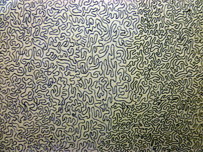
This final image is of a stomatal imprint of the topside of a spinach leaf. It reminded me of a pattern I draw a lot:
(Image: my own)
This consists of only one line. I've been filling pages with pattern for years, but haven't considered how similar it looks to cell structures. I'm aiming to use this kind of intricacy and detail within the work I produce for this project.


No comments:
Post a Comment